
Subclass-specific Digestion of IgA in Serum using IgaSAP™
Application
Subclass selective digestion of IgA in complex samples like serum, enabling targeted analysis of IgA1 and IgA2m1 without off-target cleavage.

IgA constitutes around 15% of the total amount of immunoglobulins in normal human serum. The ability to selectively digest IgA1 and IgA2m1 can be of great value in research and diagnostics as well as in bioprocessing and therapeutic applications.
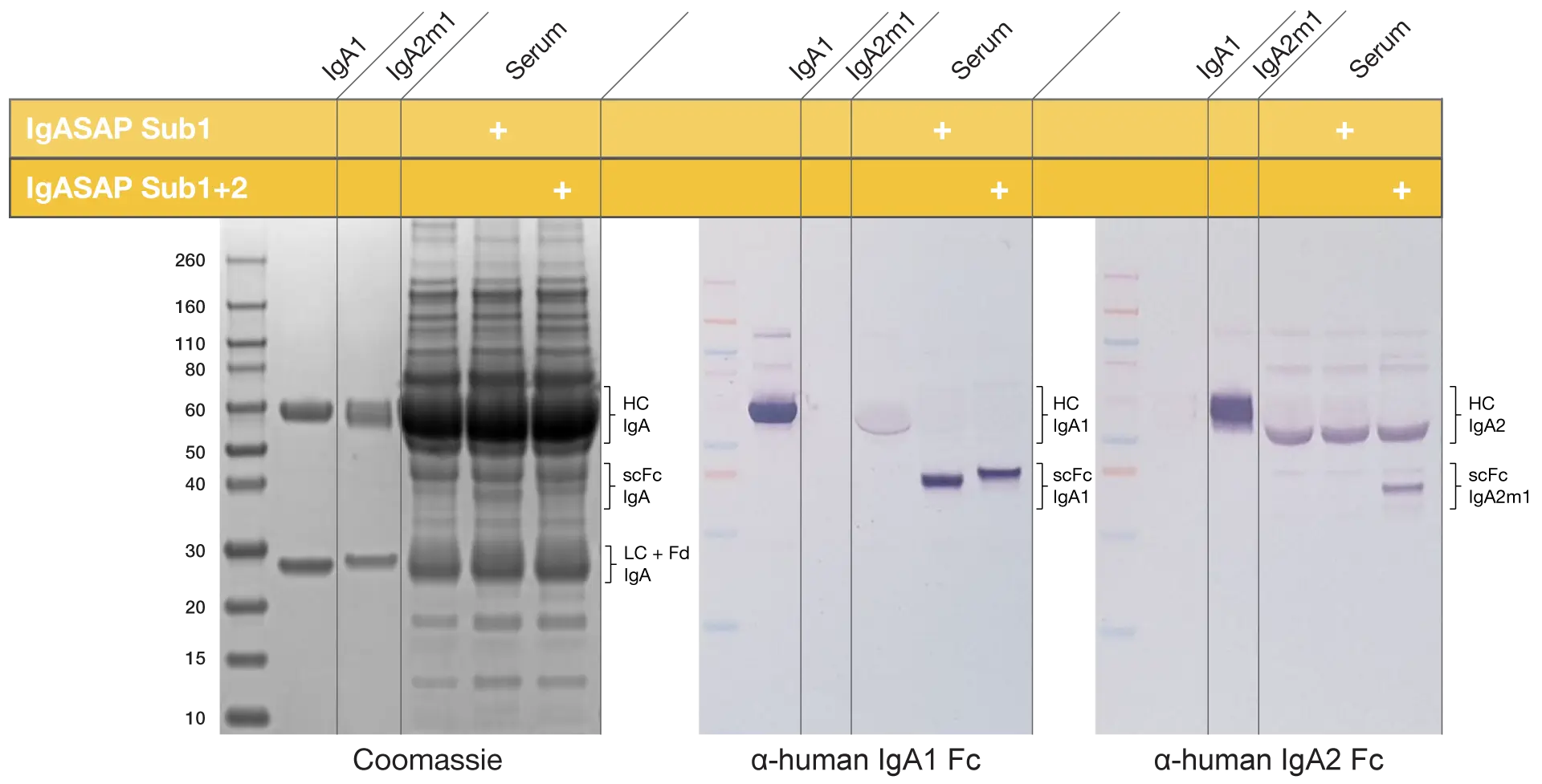
The specificity of the IgASAP enzymes make them excellent tools for such isotype-specific digestion of IgA in human serum samples or other complex sample matrices. Here, human serum was incubated with IgASAP Sub1 Lyophilized for 1 hour at 37°C and IgASAP Sub1+2 Lyophilized overnight at 37°C, before analysis with reducing SDS-PAGE and western blot.
IgASAP enzymes are excellent tools for IgA digestion in serum
IgASAP Sub1 specifically digests IgA1 in serum while leaving IgA2 and the other components of the serum intact. IgASAP Sub1+2 elicits a higher degree of digestion in the serum samples as the IgA2m1 fraction is also hydrolyzed. Remaining intact IgA2 in the pooled human serum after IgASAP Sub1+2 treatment is from the digestion-resistant allelic forms IgA2m2 and IgA2m3.
Subclass specific digestion of IgA using IgASAP Sub1 and IgASAP Sub1+2
This shows the high subclass specificity of the IgASAP enzymes and demonstrates the possibility to use the products in complex biological samples such as serum and plasma without the risk of off-target digestion.
Efficient digestion in complex matrices without the risk of off-target digestion
Related Products


Resources
IgASAP™
Download Product Folder

