
Site-specific Digestion of Human IgA1
Site-specific digestion of human IgA1, generating homogeneous Fab and Fc fragments for high-resolution middle-level LC-MS characterization.

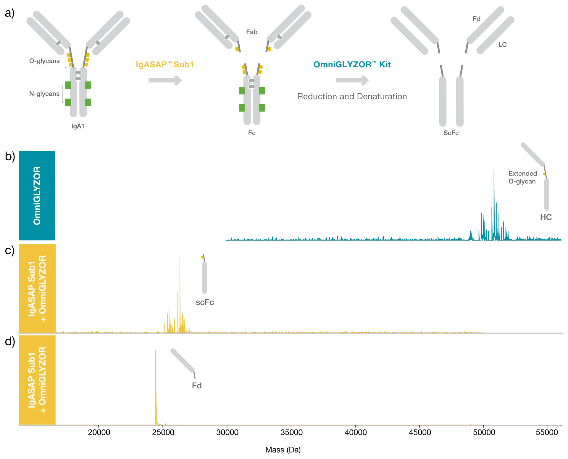
IgASAP Sub1 digests human IgA1 at one specific site above the hinge, generating Fab and Fc fragments, which allows for middle-level analysis of IgA1. To demonstrate the site-specificity of IgASAP Sub1, a commercially available human myeloma IgA1 was digested with IgASAP Sub1 Lyophilized for 1 hour at 37°C and compared to undigested IgA1.
As IgA1 carries multiple N- and O-glycans, the samples were treated with OmniGLYZOR Kit, including the additional deglycosylation step under denaturing conditions using PNGase F Lyophilized to remove all N-glycans, to facilitate the data interpretation. The samples were analyzed with LC-MS. The digestion using IgASAP Sub1 is fast, easy, and carried out under physiological conditions, and the specificity of the enzyme prevents overdigestion of IgA1.
Highly specific digestion of human IgA1 in just 1 hour
Deconvoluted mass spectra of N- and O-deglycosylated heavy chain (HC) of the IgA1 from human myeloma reveals a highly heterogenous material. After digestion using IgASAP Sub1, the heterogeneity of the HC material follows to the digested Fc part. It derives from several C-terminal truncations and a remaining extended O-glycan, not removed by OmniGLYZOR. The Fd fragment is homogenous and the acquired masses confirms the single digestion site at VPSTPP / TPSPST (aa 108/109 of constant heavy chain acc. to Uniprot p01876).
Domain-specific identification of IgA1 modifications
Despite the heterogenous starting material, the data clearly shows the single digestion site in the upper hinge region of human IgA1 and emphasizes the advantages of using a middle-level workflow to generate high resolution mass data for such complex samples.
Middle-level analysis improves analysis of human IgA1
Related Products

Resources
IgASAP™
Download Product Folder

