Fluorescent Imaging using GlyCLICK®, an Interview with ImaGene-iT
Bo Holmqvist at ImaGene-iT gives us insight about microscopy imaging and how ImaGene-iT is using the site-specific GlyCLICK® technology to obtain high-quality fluorescence images.
Tell us about yourself and ImaGene-iT?
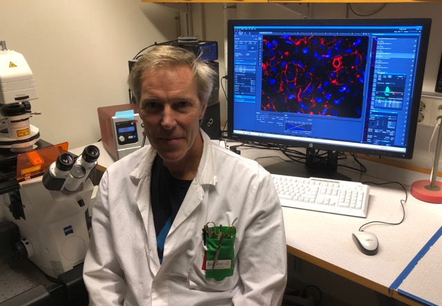
I am CSO at ImaGene-iT, an independent contract research company supporting the life science industry and academia. Concurrently, as associate professor in experimental pathology, I have a research background in histology and neurobiology with extensive experience in microscope imaging and digital image analysis. As a principal investigator at ImaGene-iT, I perform most of the quality assessments using high-resolution confocal microscopy along with imaging for further digital analysis.
Our projects comprise a variety of questions and involve various types of biological samples on both the histological and cellular levels. At ImaGene-iT, we are engaged in finding suitable solutions and strategies for our customers and partners, concerning the choice of experimental labeling techniques combined with the optimal microscope and image analysis technique.
What are the key aspects of experimental design in imaging experiments?
From the question at hand and to obtain the best end result, we want to participate in the whole chain of tailor-made procedures. This includes the initial handling of samples, the choice of labeling technology and the best suited detection and imaging equipment. This allows us to extract the most relevant data possible for high quality image analysis.
What are the advantages of using the GlyCLICK technology?
In our tests, antibodies with GlyCLICK conjugated fluorophores work very well for imaging in both in vitro cell and tissue analyses. The conjugates provide an excellent signal-to-noise ratio for detection and imaging with fluorescence microscopy. For confocal microscope analysis, GlyCLICK conjugated antibodies give an optimal range of intensity levels. This allows us to obtain the ideal settings for detection of both the lower and higher intensity levels with a reduced loss of signal and saturation. The optimal range of detected intensity levels improves the quality of assessment as well as the digital imaging which in turn improves the image analyses.
What does the future of imaging look like?
In our field, one of the major recent advances in imaging is the ability to extract a large amount of data from images, for example from multi-fluorescence labeled samples. The use of markers with a known number of conjugation sites per target molecule, such as the GlyCLICK technology offers, can significantly improve quantitation possibilities for all types of imaging-based analysis. This opportunity benefits the fields of basic research and drug development and may form the basis for exciting diagnostic tools in clinical histopathology. The combined use of GlyCLICK conjugated fluorescence with small animal in vivo imaging could further expand the quality of quantitative data that can be collected for our clients, from the cellular level to the whole animal.
Read more about the Genovis GlyCLICK technology and its applications.
![]()