
Site-specific Digestion of IgM
Site-specific IgM digestion under physiological conditions, producing homogeneous fragments for precise middle-level LC-MS characterization.

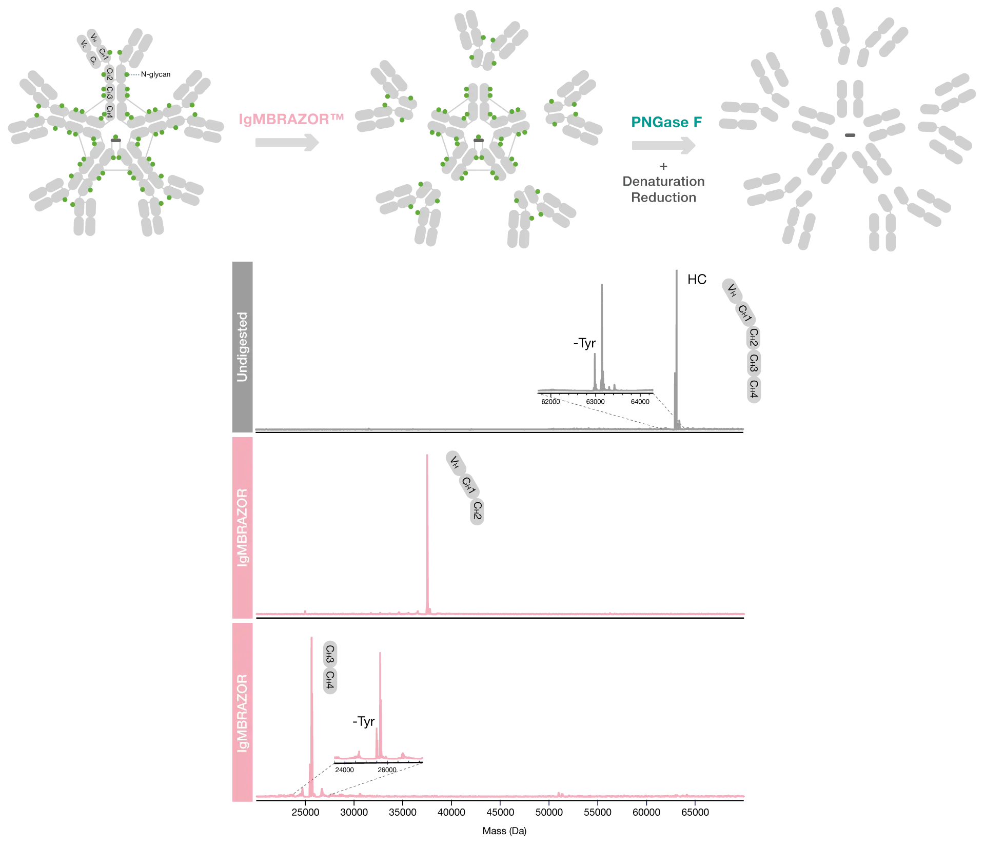
To demonstrate the site-specificity of IgMBRAZOR, human myeloma IgM was digested with IgMBRAZOR Lyophilized for 30 minutes at 37°C and compared to undigested IgM. To facilitate data interpretation, the N-glycans were removed using PNGase F under denaturing and reducing conditions. The homogenous fragments observed in the mass spectra after IgMBRAZOR and PNGase F digestion and the masses of the detected fragments were consistent with digestion at one single site (..VPDQDT / AIRVFA..).
Highly specific digestion of IgM at one single site
The digestion using IgMBRAZOR is fast, easy, and carried out under physiological conditions, and the specificity of the enzyme prevents overdigestion of IgM and hydrolysis of other proteins, challenges otherwise commonly associated with other proteolytic enzymes.
Fast and efficient digestion with no risk of overdigestion
Middle-level analysis allows for the generation of high resolution mass data with minimal sample preparation and data analysis, which can be efficiently used for characterization, quality control, stability testing and production monitoring of therapeutic antibodies.
Simplified characterization IgM using middle-level analysis
Related Products

Resources
IgMBRAZOR™
Download Product Folder

