
Deglycosylation of the C1 inhibitor using GalNAcEXO®
Application
Confirmation and quantification of Tn antigens on O-glycosylated biopharmaceuticals while simplifying intact-mass analysis and revealing protein variants.

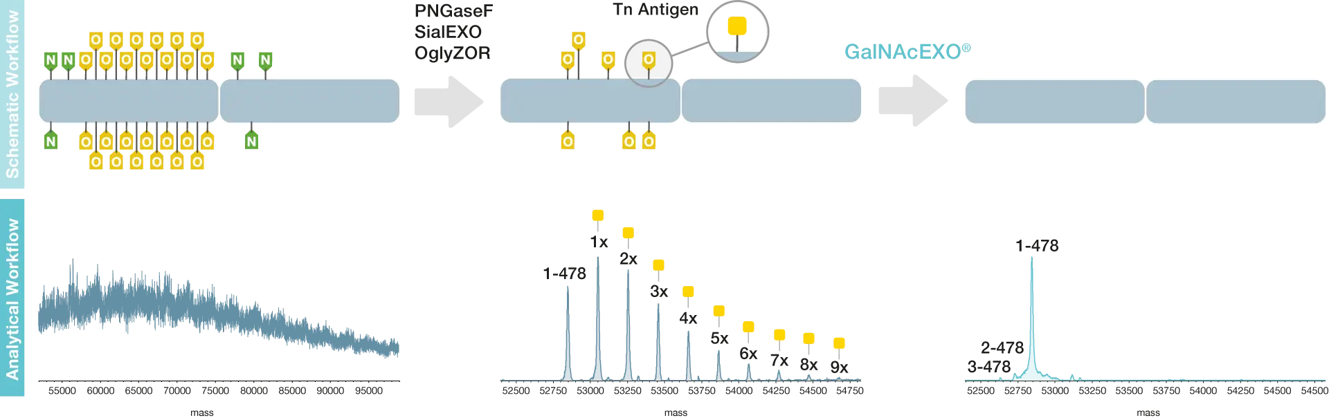
During production of O-glycosylated biopharmaceuticals, Tn antigens may appear as a result of incomplete processing of the core GalNAc residue. This appears as a repeating mass shifts with a 203 Da HexNAc unit difference.
To confirm the presence of the Tn antigen, the following workflow can be applied, here demonstrated on the heavily (26 sites) O-glycosylated C1 inhibitor. First, the glycoprotein is trimmed of N-glycans using PNGaseF and core 1 O-glycans using OglyZOR and SialEXO. A pattern of repeating peaks can be observed in the mass spectra. Secondly, after incubation with the GalNAcEXO enzyme, the remaining GalNAc residues were completely removed leaving one single peak. This workflow therefore confirms the presence of Tn antigens on O-glycosylated biopharmaceuticals.
Confirmation of Tn-antigen presence by digestion using GalNAcEXO
Partial deglycosylation of the human C1 inhibitor allows for quantification of the number of Tn-antigens present on the molecule. This is important for process development and comparability studies, and quantification of this kind is not possible when the fully intact glycans are present on the molecule.
Quantificaiton of Tn-antigen population on complex glycoproteins
Reduction of the remaining heterogeneity enables more confident confirmation of the intact mass of the protein. Complete removal of the glycans also makes it much simpler to identify any protein variants that may be present. This is demonstrated through the identification of some truncated variants of the C1protein following removal of the Tn-antigen using GalNAcEXO.
Efficient Tn-antigen hydrolysis reduces heterogeneity to allow identification of underlying protein variants
Related Products

Resources
GalNAcEXO®
Download Product Folder

SmartEnzymes™ for the Glycosylation Charcterization of Glycoproteins
Download Scientific Poster


