SmartEnzymes™ for AAV Gene Therapy Applications
Download Scientific Poster


Application
Efficient IgG cleavage with a low immunogenicity risk enzyme, supporting clearance of neutralizing antibodies to improve gene therapy administration.

Neutralizing anti-AAV antibodies are common in the human population and can exclude patients from lifesaving treatments. A risk for treatment is that potential re-dosing of the gene therapies may increase the formation of neutralizing antibodies against the vector. Strategies to overcome neutralizing antibodies include plasmaphoresis, direct organ delivery, engineered AAVs or immunosuppression.
Clearance of neutralizing antibodies by IgG-specific digestion is a potential strategy to increase addressable patient population for gene therapies. IdeS, IdeZ and Xork are IgG-specific proteases against human IgG, but the enzyme sequences are very different. IdeS and IdeZ have been discovered in common bacterial pathogens and most human subjects have encountered IdeS through streptococcal infections, and have anti-IdeS antibodies in their bloodstream. Xork is a novel enzyme with low sequence identity to IdeS and IdeZ, and is not derived from a human pathogen. Using an enzyme derived from a non-human pathogen may reduce the risk of having neutralizing antibodies against the enzyme itself. In this work, immunosuppression using Xork as a potential tool to facilitate neutralizing antibody clearance is explored.
Xork as a tool to improve gene therapy with reduced risk of anti-enzyme antibodies
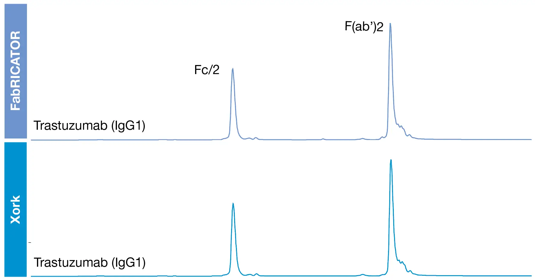
The Xork protease has a low sequence identity to IdeS and the enzymes’ digestion was compared were compared using standard IgG. The digestion patterns were analyzed using RP-LC. The data confirm that Xork and IdeS rapidly digest human IgG into F(ab’)2 and Fc fragments (Fig. 1). The digestion site of Xork was determined to be located below the hinge of IgG using a human IgG1 antibody as reference. After digestion, reduction, and LC-MS analysis, the masses of the subunits Fd’, LC and Fc/2 confirmed the digestion site between the glycine residues in the hinge region.
Efficient digestion of human IgG using Xork
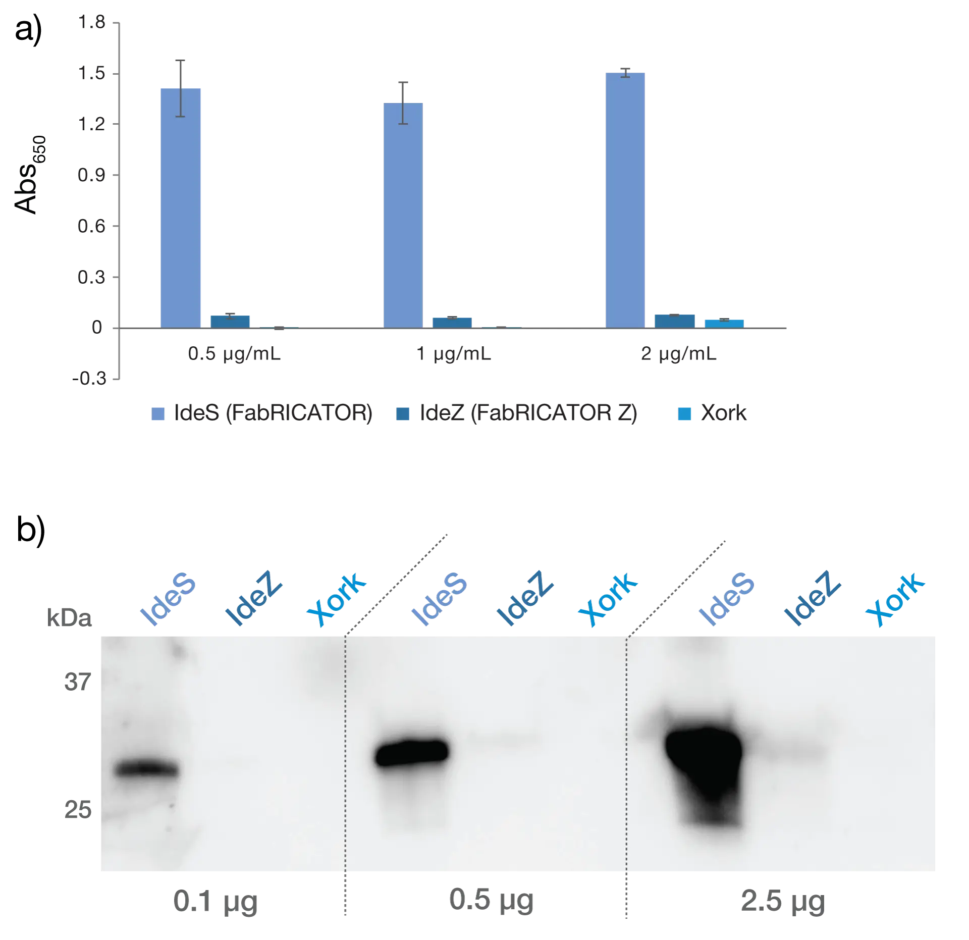
The presence of antibodies targeting the bacterial enzymes was studied by ELISA and western blot (Fig. 2). As detection, pooled human immunoglobulins (IVIG) was used. The strong signal from IdeS demonstrates the common presence of antibodies targeting this enzyme. The lower signals from IdeZ and Xork reflect that sources of those enzymes are bacteria not infecting humans, thus no or a limited amount of antibodies are present in IVIG. The combination of high efficiency towards human IgG and no detectable anti-enzyme antibodies in IVIG demonstrates the high potential of Xork as an effective tool to facilitate gene therapy treatments.
No detection of anti-Xork antibodies in IVIG
Download Scientific Poster
Download Product Folder
