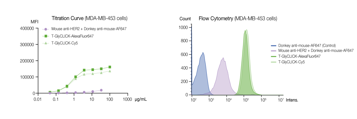
Flow Cytometry
Flow Cytometry
Labeled antibodies offer the ability to quantify distinct markers or cell populations using flow cytometry in cell sorting, imaging or immunophenotyping experiments. Direct detection using GlyCLICK labeled antibodies preserves the sensitivity of the analysis while minimizing both protocol complexity and unspecific binding. Here, we demonstrate the GlyCLICK technology and its advantages in sensitive or multiplexed flow cytometry analysis.
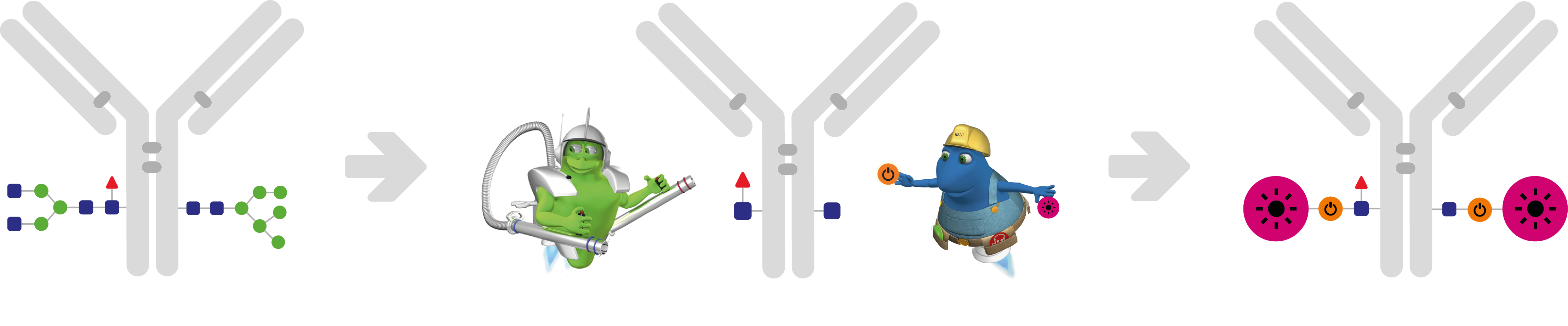
Preserved Immunoreactivity with GlyCLICK
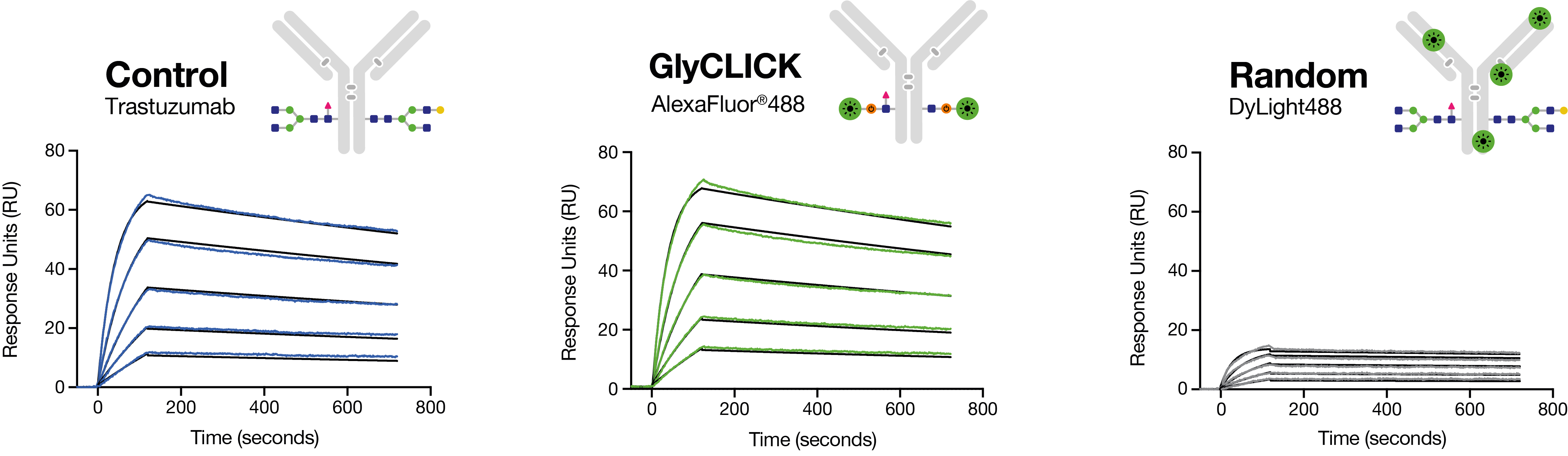
Randomly labeled antibodies can impact the quality of the analysis through unspecific binding and inadequate signals due to excessive labeling and impaired immunoreactivity. To evaluate the antigen-binding capacity after labeling, trastuzumab was site-specifically conjugated to AlexaFluor®488 using GlyCLICK or by random labeling at lysines using DyLight®488. The resulting response curves show that only 20% of the randomly labeled material retained binding capacity while GlyCLICK conjugated antibodies display full preserved immunoreactivity for sensitive and reliable detection (Fig. 1).
Increased Flexibility for Multiplexed Analyses
The desired combination of antibody and label may not be available or suffer from poor conjugation quality that impacts the sensitivity. GlyCLICK labels native IgG specifically at the Fc glycan sites, incorporating precisely two labels (DOL=2.0) and abolishing interactions with Fc receptors, the effector functions. To demonstrate the site-specific GlyCLICK conjugation, trastuzumab was labeled and analyzed at subunit level using FabRICATOR and LC-MS. The resulting mass spectra show that all native Fc-glycoforms (Fig. 2a) are trimmed as indicated by the mass shifts corresponding to deglycosylated Fc/2 (Fig. 2b). The exposed Fc glycan site is then azide activated (Fig. 2c) for labeling using click chemistry and the alkyne-carrying fluorophore of choice (Fig. 2d, 2e).
Figure 2. Deconvoluted mass spectra of trastuzumab Fc/2 fragments after FabRICATOR digestion and reduction showing a) native trastuzumab, b) trastuzumab Fc/2 deglycosylated to the inner GlcNAc by the GlycINATOR enzyme , c) azide activated Fc/2 fragments, d) T-GlyCLICK-AlexaFluor®647 and e) T-GlyCLICK-Cy5.
Sensitive Detection of Biomarkers
Multiplexing can result in unwanted fluorescence signals and noise levels originating from unspecific staining, elevated background and spillover that reduces both the sensitivity and resolution of the analysis. The ability to discriminate between negative and positive populations of cells can thus be affected, and a low separation index is often observed. The performance of site-specifically labeled antibodies conjugated using GlyCLICK was evaluated for flow cytometry imaging and compared to an indirect detection method.
HER2 positive cells were immunostained with T-GlyCLICK-AlexaFluor®647 or T-GlyCLICK-Cy5 and compared to indirect detection using primary and secondary antibodies. To minimize unspecific binding, titration was determined the saturation level of 3.7 ug/ml (Fig. 3a). The results show both higher intensities and a better separation of negative and positive populations with a 10-fold increase in separation index using GlyCLICK conjugated antibodies (Fig. 3b).
Figure 3. Fluorescent imaging of high expressing HER2-positive cells showing a) titration curve of MDA-MB-453 cells against antibody in solution 1:200 (0.046-100 µg/ml), b) flow cytometry analysis showing fluorescence intensity and count of MDA-MB-453 cells either unstained with donkey anti-mouse-AF647 (control) or stained with mouse anti-HER2 + donkey anti-mouse-AF647, T-GlyCLICK-AlexaFluor®647 or T-GlyCLICK-Cy5.