Sugar-free Proteins in a Few Hours
OmniGLYZOR Hydrolyzes N- and Mucin-type O-glycans
Glycosylation tends to be heterogeneous, making analysis of heavily glycosylated biotherapeutics challenging. Confirmation of intact mass as well as characterization of other product quality attributes are hampered by the complexity of the different glycoforms which is why efficient tools for glycan removal greatly simplify such analyses.
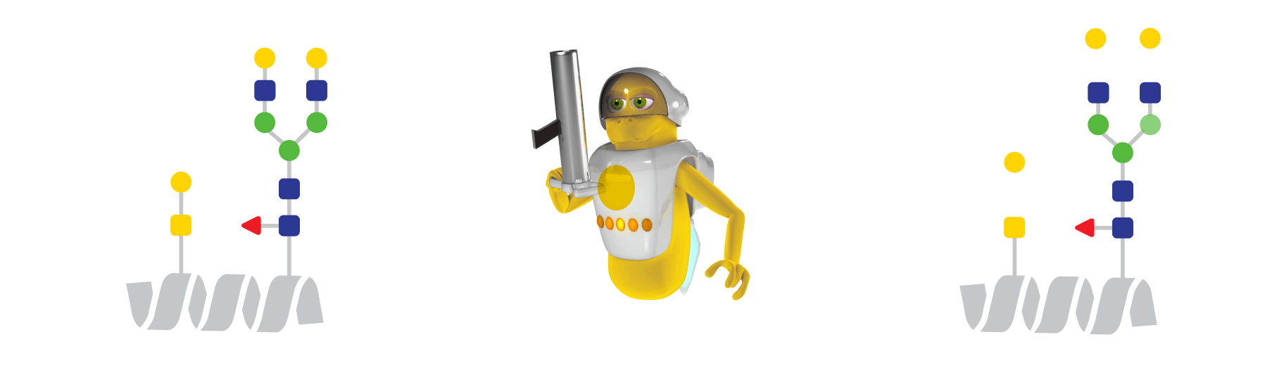
While N-glycans contain a common core structure and can be removed en bloc by PNGase F, mucin-type O-glycans are more structurally diverse with several different cores. OmniGLYZOR contains a mixture of immobilized enzymes able to sequentially break down the O-glycans for the complete removal of the most commonly occurring O-glycan structures (di- and mono-sialyl core 1 and Tn antigen).


Efficient Removal of N- and Mucin-type O-glycans
Here we demonstrate the performance of OmniGLYZOR using two therapeutic proteins as substrates. Etanercept is an Fc-fusion protein containing the extracellular domain of the TNFα receptor. This domain is modified with 2 N-glycans and 8-11 core 1 O-glycans with varying degrees of sialylation. Incubation of this heavily glycosylated protein on OmniGLYZOR Microspin columns for 1 hour at 37°C led to the complete removal of all N- and O-glycans as demonstrated by middle-level LC-MS analysis (Fig 1a). Erythropoietin (EPO) is a small protein carrying 3 N-glycans and 1 core 1 O-glycan resulting in close to 40% of its total mass consists of glycans. When analyzed in its intact state, the glycan heterogeneity of EPO results in a very complex mass spectrum. By incubating on OmniGLYZOR Microspin columns for 1 hour at 37°C, the N-and O-glycans are efficiently removed as indicated by a single peak corresponding to the unmodified protein (Fig 1b). A minor amount of O-glycans modified with acetylated sialic acids is left on EPO, since such structures are inefficiently hydrolyzed by OmniGLYZOR. Both substrate proteins were also treated with another commercially available deglycosylation product according to the manufacturer’s recommendation (O/N incubation at 37°C) and the data are shown for comparison.

Figure 1. Complete hydrolysis of N- and O-glycans by OmniGLYZOR. Deglycosylation of a) etanercept and b) EPO. The substrate proteins were incubated on OmniGLYZOR Microspin columns for 1 h at 37°C, or with another commercially available deglycosylation product for 1 h and 20 h at 37°C. To simplify the analysis of etanercept, the deglycosylated protein was digested with FabRICATOR to separate the O-glycosylated TNFR domain from the Fc fragment and the resulting subunits were analyzed by reversed- phase LC-MS using a Waters™ BioAccord™ LC-MS system. EPO was analyzed in the same way in its intact state. The peaks corresponding to the fully deglycosylated substrate proteins are shaded in orange.
Deglycosylation under Denaturing Reaction Conditions

Figure 2. Complete deglycosylation of plasma-derived human C1 inhibitor. The protein was analyzed by reversed-phase LC-MS using a Waters™ BioAccord™ LC-MS system in its untreated state (top), after deglycosylation on an OmniGLYZOR Microspin column under native conditions (middle) and after additional deglycosylation using PNGase F under denaturing conditions (bottom). The peak corresponding to the deglycosylated substrate protein is shaded in orange.
O-linked glycans are generally found at exposed locations on the protein as they are attached after protein folding has occurred. Only such exposed sites are accessible to the glycosyltransferases responsible for their synthesis. N-glycans, on the other hand, are attached co-translationally to the unfolded protein in the ER. This results in certain N-glycosylation sites being poorly accessible to PNGase F or not accessible at all once the substrate protein is folded into its native state. Hydrolysis of such N-glycans therefore requires the substrate protein to be denatured in a way that is compatible with enzymatic activity.
Here we analyzed the C1-inhibitor – a human plasma-derived biotherapeutic modified with 6 N-glycans and up to 28 O-glycans consisting predominantly of sialyl core 1 structures. Without any pretreatment of this highly heterogeneous protein, reversed-phase LC-MS analysis yielded a complex mass spectrum impossible to interpret in detail (Fig. 2). Using OmniGLYZOR Microspin columns under native conditions, all O-glycans were efficiently removed within 3 hours. However, between 1 and 3 N-glycans remained on the protein. These inaccessible N-glycans were removed by an additional deglycosylation step under denaturing conditions using the lyophilized PNGase F and the MS-friendly RapiGest™ SF surfactant* included in the OmniGLYZOR kit. Complete removal of all glycans was observed, with the exception of the minor amount of core 2 O-glycans present on the molecule. The enzymes hydrolyzing the O-glycans do not benefit from denaturation, which is why the OmniGLYZOR Microspin columns should only be used under native conditions.
* RapiGest™ SF Surfactant from Waters Corporation is included in OmniGLYZOR. RapiGest™ is a trademark of Waters Corporation.
Learn more
Popular FAQ
No, both N- and O-glycans are trimmed by the exoglycosidases present in OmniGLYZOR and do not longer represent the structures found on the intact glycoprotein substrate.
No, it is not recommended. We can only guarantee optimal performance for one-time use.
OmniGLYZOR hydrolyzes the amide bond between the polypeptide asparagine and the innermost GlcNAc of all mammalian asparagine-linked complex, hybrid, or high mannose oligosaccharides. It does not remove N-glycans with α1-3 core fucosylation.